Musculoskeletal scans
A Musculoskeletal scan is an examination of a musculoskeletal structure: soft tissue lumps, muscle, tendons and joints.
- A 20 minute scan;
- A written ultrasound report of the examination immediately after the scan.
We’ll see you in no time
Some of our clients choose to book an ultrasound scan with us after having been referred for a musculoskeletal scan in the NHS by their GP. Due to long waiting times they are unwilling to wait for the scan. Going privately allows them to have quicker and more convenient appointments with faster results.
Ultrasound does not involve radiation and is in real time i.e. live imaging and is not static, CT involves radiation. Ultrasound can be used to scans many parts of the body.
As with many tests, ultrasound scans have limitations such as unable to visualise bone, or inside the joint space within the shoulder or knee therefore may not be suitable in certain situations and like any test cannot diagnose every problem. Please contact us if you are not sure if this is suitable for you.
Helping multiple diagnoses
Ultrasound is typically used to help diagnose:
- Tendon tears, or tendinitis of the rotator cuff in the shoulder, Achilles tendon in the ankle and other tendons throughout the body;
- Muscle tears, masses or fluid collections;
- Ligament sprains or tears
- Inflammation or fluid (effusions) within the bursae and joints;
- Benign and malignant soft tissue tumours;
- Ganglion cysts;
- Hernias;
- Foreign bodies in the soft tissues (such as splinters or glass);
- Soft tissue lumps.
The ultrasound scanning method
Ultrasound gel is placed on the anatomy to be scanned the areas of interest are scanned with an ultrasound probe. Multiple images of the area scanned are taken; you may need to move your shoulder/elbow/hand/hip/knee/foot in different positions to enable us to see the anatomy clearer. The results of the scan will be given to you immediately after the scan.
Colour Doppler technique
Colour Doppler techniques can be used to measure neovascularization within the synovial lining of the joint, tendons, and soft-tissue masses.
Why try ultrasound?
There are many kinds of ultrasound scans that can be performed as ultrasound has numerous applications in healthcare and are ultimately beneficial to us if used in the right context by a competent body or person. However as with any test, ultrasound scans have limitations some of which are generic to all scans such as not being able to visualise bone clearly or scan specific and therefore may not be suitable in certain situations and like any test cannot diagnose every problem.
Preparation
No preparation is required – you only need to expose the area to be scanned.
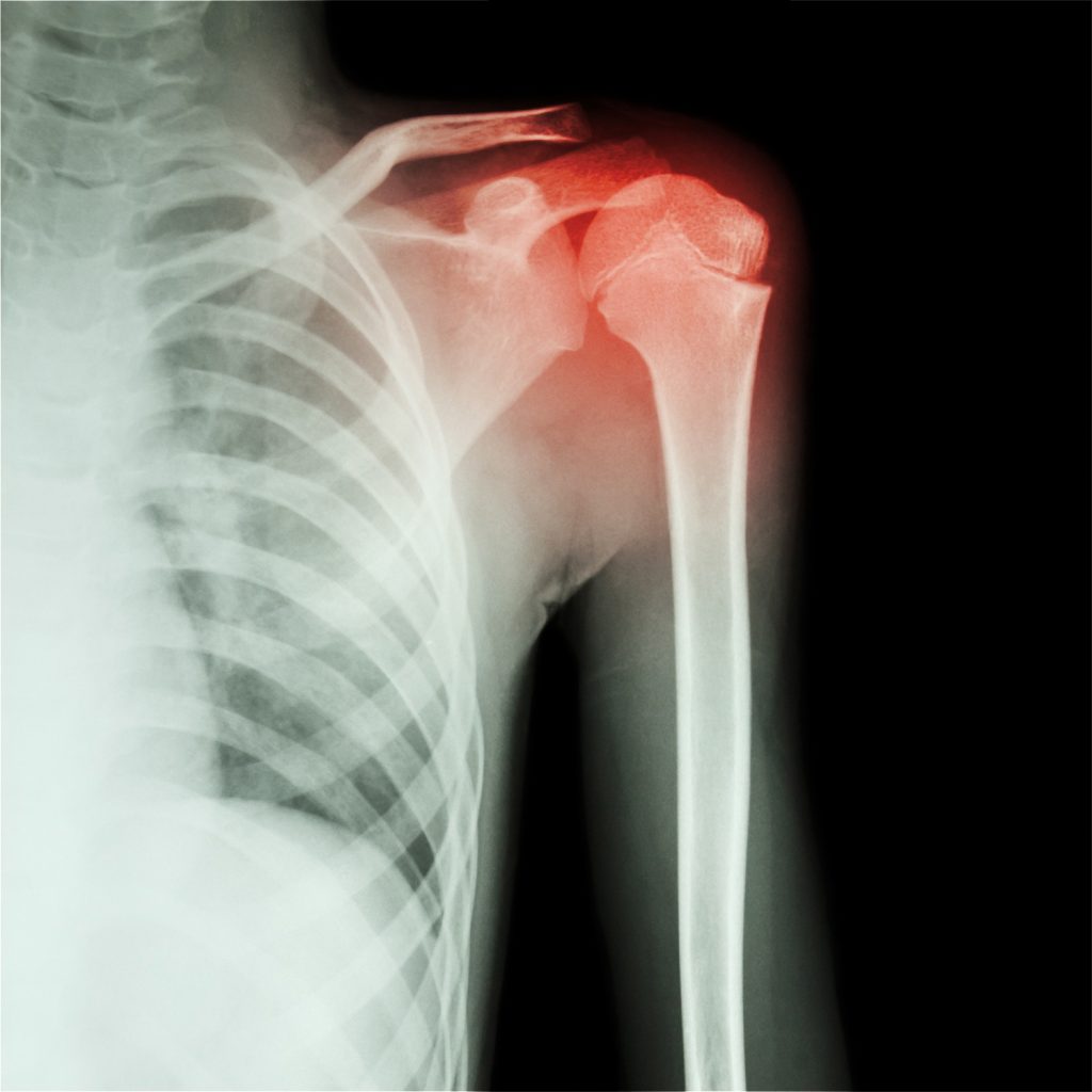
Musculoskeletal shoulder
Shoulder examination
The shoulder and rotator cuff are examined: we will assess the joint, rotator cuff tendons: (the biceps tendon and sheath, subscapularis, supraspinatus and infraspinatus tendon), muscles, ligaments, bursa and any soft-tissue swellings. The rotator cuff holds the shoulder joint together. Problems with the rotator cuff tendons can be the cause of many shoulder problems, which are becoming increasingly common due to the stress and strains.
The acromioclavicular joint is also examined.
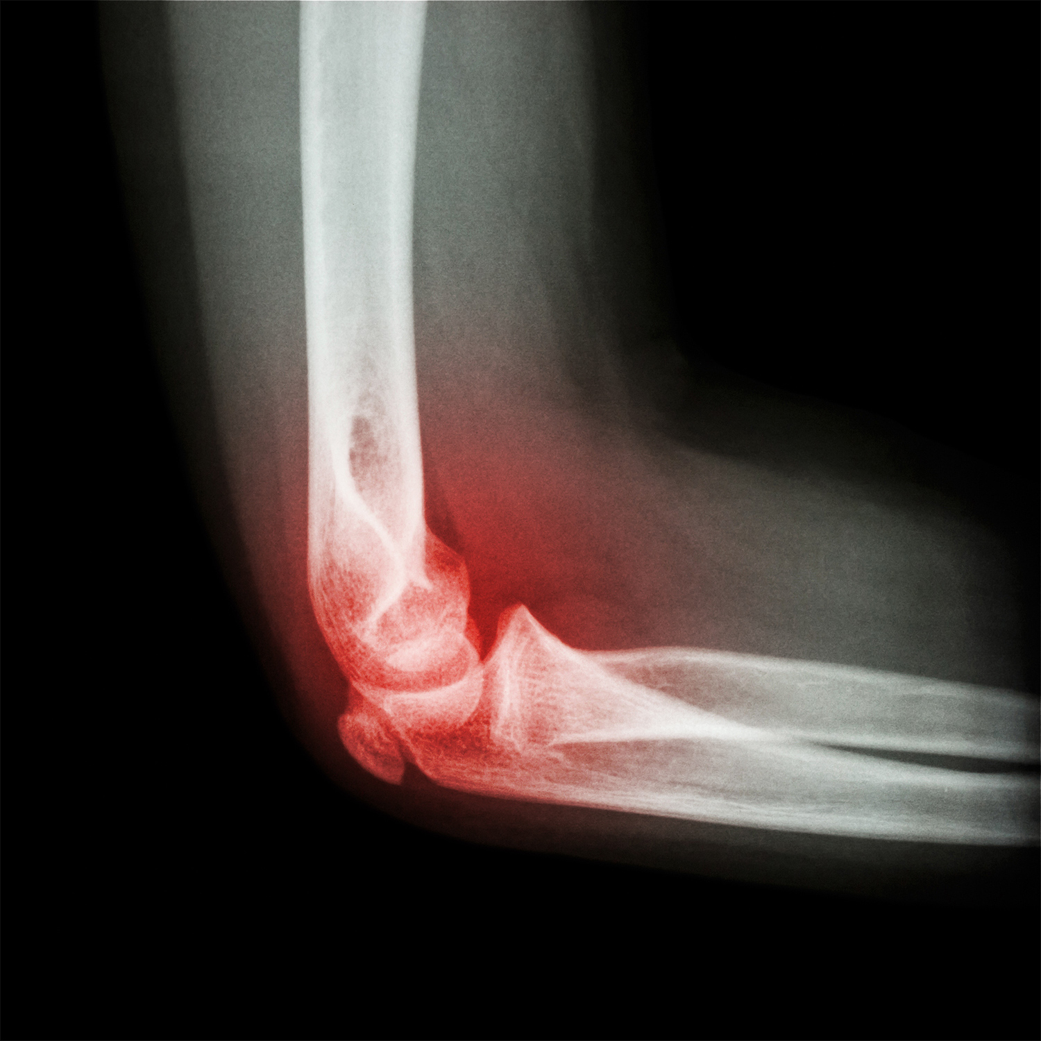
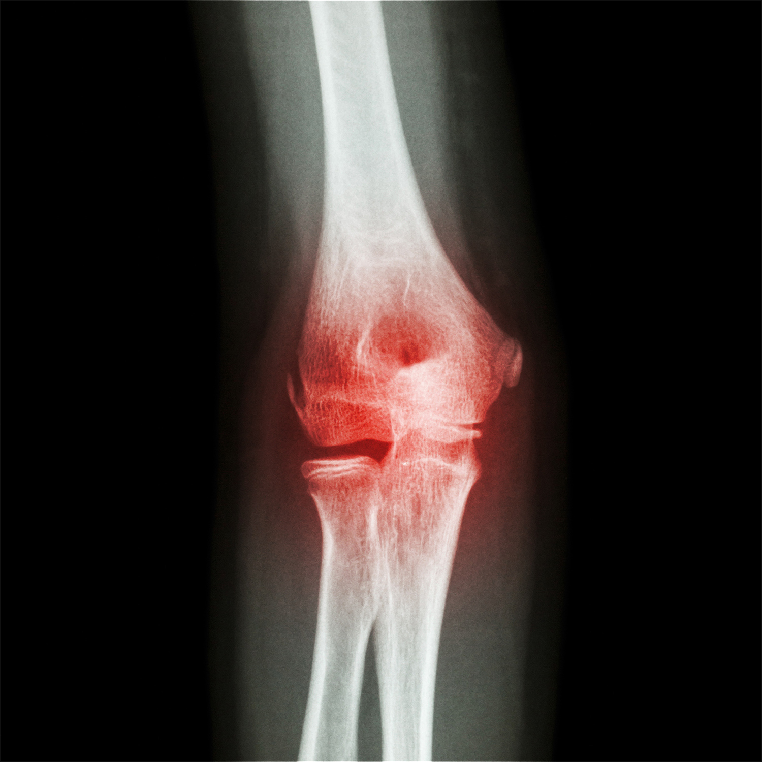
Musculoskeletal elbow and forearm
Elbow and forearm scan
Ultrasound scan of the elbow can look at the joint, the tendons, muscles, and ligaments. It also looks for bursa and soft-tissue swelling.
Commonly seen due to pain in the elbow on either side of the elbow (known as tennis elbow or golfer’s elbow) is due to inflammation of the tendons. Ultra nerve neuropathy can also be visualised.
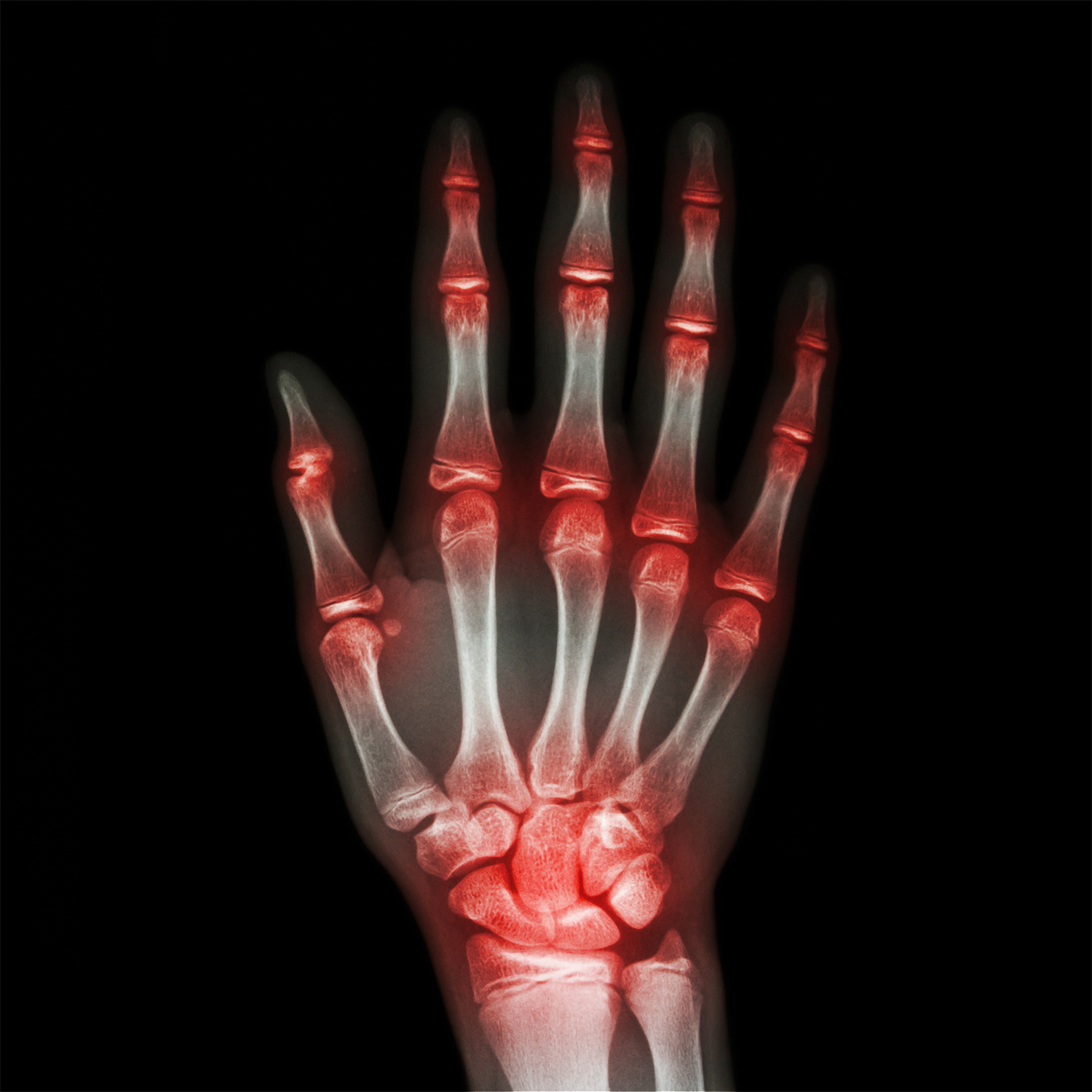
Musculoskeletal hand and wrist
Hand and wrist scans
Tendon inflammation and rupture can be visualised, also lumps and bumps, even glass or thorn (foreign bodies) can be seen within the finger.
Hand and wrist injuries are also common and can affect ligaments, joint surfaces, tendons, muscles and nerves.
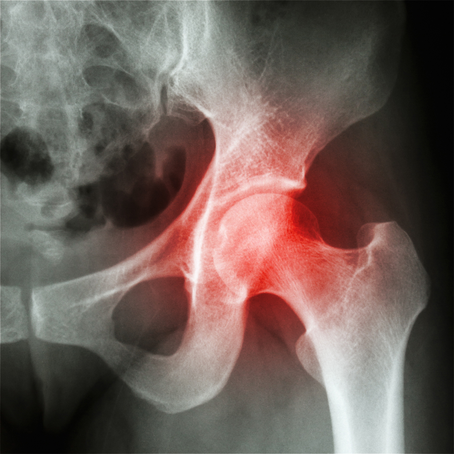
Musculoskeletal hip and groin
Hip and groin scans
Hip and groin pain are very common and ultrasound is very good at assessing the hip tendons, ligaments, muscles, nerves, synovial recesses, articular cartilage, bone surfaces and joint capsule.
With ultrasound, we scan is to detect and localize pathological processes, to differentiate between intra articular and extra articular pathology.
Many hip diseases are detectable with ultrasound including assessment of the soft tissues, tendons, ligaments and muscles, and also of the bone structures and joint spaces.
Hernias can also be excluded in the groin such as inguinal hernias and femoral hernias, spigelian hernias and anterior abdominal wall hernias.
Musculoskeletal knee
Knee scan
The knee joint is made up bones, cartilage, ligaments, and tendons.
Ultrasound scan provide useful information on a wide range of conditions affecting parts of the knee, such as the tendons, ligaments, muscles, synovial space, articular cartilage, and surrounding soft tissues.
It can also exclude/confirm baker’s cyst at the back of the knee.
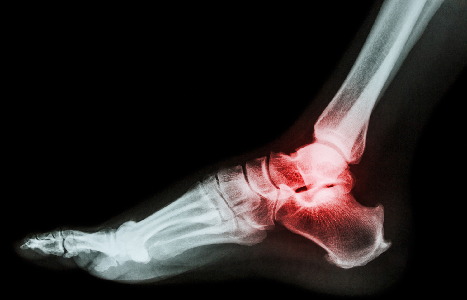
Musculoskeletal ankle and foot
Foot and ankle scans
The ankle and foot has many tendons, ligaments and bones, pain or an injury are common causes to investigate the causes for inflammation of the tendons, bursitis, degeneraltive changes such as arthritis, and joint effusions.
Lumps can be investigated such as ganglions, neuromas and fibromas.
Painful heels can be scan to exclude/confirm plantar fasciitis.
Pricing
| Service | Price |
|---|---|
| Shoulder scan | £160 |
| Elbow scan | £130 |
| Hand/Wrist scan | £130 |
| Hip scan | £150 |
| Knee scan | £130 |
| Foot/Ankle scan | £130 |